
In order to understand what you have and why, you need to understand a little bit about how veins work. Think of venous anatomy as an interstate highway system. You have highways coming and going, twisting and turning, and these routes or tributaries serve different destinations. These destinations fulfill our need to arrive at work on time. They fuel our passion for exploration or relaxation. They allow us to move freely across vast regions. Our highways and roads lead over bridges, through tunnels, and often are unfamiliar territory to us. When something is unfamiliar to us, we get nervous, uptight, or discount help from others. That is why men don’t ask for directions. Our goal at the Center for Venous Disease is to get you familiar with the twisting and turning of the interstate that lies within your venous and vascular system and help offer solutions and directions.
Just like our highways, there are different routes to take and different reasons we take the road we’re on. In our legs, there are a few main veins mentioned. They are:
- Common Femoral Vein
- Femoral Vein
- Great Saphenous Vein (GSV)
- Small Saphenous Vein (SSV)
- Sural Veins
- Mid Thigh Perforators (PERFs)
- Paratibial Perforators
- Posterior Tibial Perforators
- Intergemellar Perforators
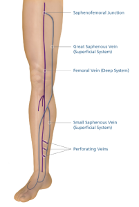
Picture provided by Covidien.
Side note: The GSV, SSV, and Perforator Veins are the most common veins for ablation procedures for treatment of Varicose Veins.
Where your Common Femoral Artery, Common Femoral Vein, and Great Saphenous Vein come together in the groin is generally known as the Saphenofemoral Junction. A photo of this image on ultrasound is below. We often refer to this as the “Mickey Mouse” view with patients because it kind of resembles Mickey’s ears in this view. This junction is an area of the body where high volumes of blood move down the legs and then returns to the heart. The junction lies in the crease of the groin in the pelvic area.
The superficial vein located behind the leg below knee level and in the calf region running down towards the back of your foot is the Small Saphenous Vein (SSV). The superficial system (GSV, SSV) usually lie between .05-1.0 cm from your skin, making its location close to the surface of your skin. The Femoral Vein is down farther from the skin and runs down the leg from your groin to the ankle area and connects to the GSV. Communicating veins between the Femoral Vein and the Great Saphenous Vein or Small Saphenous Vein are called Perforator Veins. Perforator Veins, or PERFS, run up and down the entire leg and act as bridges between the GSV and the deep system. The above mentioned veins are all part of the highway, and all have to work together in order to work efficiently.
In the body, veins carry oxygen-depleted blood back to the heart through healthy veins. The arteries carry oxygen-rich blood away from the heart to all of your major organs and extremities. By treating the veins, no harm should come to the arteries. Veins return blood to the heart by way of the heart pump and the calf muscle pump that occurs when you walk or exercise. This leads to normal circulation of blood flow throughout your body. Chronic Venous Disease occurs when the blood that is supposed to be flowing back to the heart doesn’t reach the heart efficiently. The blood that doesn’t reach the heart often “pools” in the lower leg region usually in the thigh, calf level or lower to the ankle. This excessive “pooling” of blood causes leg fatigue, swelling, skin color changes (due to the iron content in the blood) and “pop outs” or “blow outs” present on your legs as Varicose Veins. Left alone, the varicose vein issues will worsen, and the skin color changes will become more apparent. The thickening of skin or a leather-like look and feel will be the next visible signs, followed by a venous ulceration or skin sore. Not only is the ulcer problematic but it is very costly. The estimated cost to treat a venous stasis ulcer is over $40,000.
A good illustration of normal valve operation and a damaged vein valve is diagrammed below:
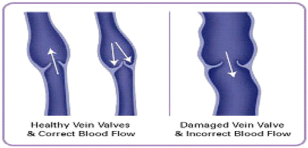
The left-hand diagram shows normal blood flow in one direction with the tiny valve closing and stopping the downward flow. The damaged valve on the right shows the valve staying open causing the blood to flow downward. This is venous reflux, referred to as an incompetent valve, which is the start of something worse. As the pressure builds, other valves fail, and the “pop outs” occur. These “pop outs” or varicose areas are a direct result of valve damage in your legs. Unless treated, the pressure will continue to increase, and more damage will occur down the leg.
A healthy vein will restore normal venous flow to the area, while a damaged vein will allow blood to flow retrograde, or downward, causing blood to pool in an area of your lower legs and ankles. If left untreated, serious problems can occur including skin breakdown, a Venous Ulcer, flaking skin, a blood clot, or all of the above.